 |
| Fig. 1 - Requirements for Fermenting Yeast |
Fermentation is a process carried out in living
cells as a means of obtaining energy from carbohydrates in the absence of
oxygen. It is essentially, therefore, a form of anaerobic respiration. In all
cases, the molecule pyruvate is formed as an intermediate product, but the
final chemicals produced depend largely on the type of organism carrying out
fermentation.
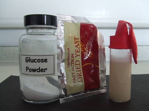
In
both wine making and bread making the microorganism involved is yeast, a
unicellular fungus. In each process the yeast converts glucose to carbon
dioxide and ethanol (see figure 1), according to the following equation:
glucose
→
ethanol + carbon dioxide
When
bread is produced the carbon dioxide is used to make the dough rise and the
ethanol evaporates in the cooking process. In wine production, however, the
carbon dioxide is allowed to escape (except in sparkling wines) while the
ethanol is retained.
Producing Ethanol - Materials and
Teaching Method
This
experiment is best suited to a Science laboratory but could be improvised
adequately in a normal classroom. Students may not be familiar with the odour
of alcohol when asked to smell each flask, but it could be suggested to them
that it is similar to methylated spirits or the smell of felt-tipped pens.
The
purpose of the cotton wool wadding is to provide a warm environment for the
yeast to multiply in the presence of glucose and water in the glucose
solution.The materials required per four students are as follows:
•
2 conical flasks fitted with a cork, glass tubing and
about 10cm attached rubber tubing
•
2 normal test tubes
•
limewater
 |
| Fig. 2 - Carbon dioxide Prodiced in Fermentation |
•
10% glucose solution (can be made by dissolving 5 glucose
tablets in 300ml water)
•
1 teaspoon dried yeast
•
Cotton wool wadding
•
Test tube rack
•
cellotape
Students
should be instructed to copy down the following directions, which should be
followed by a teacher-led explanation.
•
Add 100ml glucose solution to each conical flask.
•
Place 1 teaspoon dried yeast in one of the flasks.
•
Wrap both flasks in cotton wool and fasten with cellotape.
•
Place the cork and tubing on each flask. Submerge the free
ends of each tube in two separate test tubes containing 10 ml limewater.
•
Allow to stand for 12 hours.
The
following questions could be written on the board after students write up the
experiment and their observations.
 |
| Fig. 3 - Test for Carbon Dioxide |
1.
Describe any differences you noticed in the limewater in
each experimental set-up.
2.
In which flask did you observe foaming and bubbles?
3.
Smell the contents of each conical flask. Which one has an
alcoholic smell?
4.
Suggest why both flasks were wrapped in cotton wool.
5.
Complete this conclusion: ________ and glucose are needed
for fermentation to occur. The products of fermentation are ________ gas and
ethanol. Carbon dioxide gas turns limewater ________ (see figure 2).
Ethanol Production Follow-Up Activities
Students
could repeat the experiment, this time stretching a balloon over the mouth of
each conical flask instead of connecting the flasks to a test tube of
limewater. The flask with yeast in it should produce carbon dioxide gas, which
blows up the balloon (see figure 3).
Making Bread - Materials and Teaching Method
In
this activity a crockpot or bread making machine would both be ideal for use in
a classroom, but if neither are available the dough can be prepared in the room
and then taken to a school oven. These ingredients make one medium sized
loaf.The class could work together to make one loaf or, if multiple cookers are
available, could work in smaller groups. The materials required are:
•
Electric crockpot or bread maker
•
290 ml warmwater
•
2 tablespoons oil
•
1 1/2 teaspoons salt
•
2 tablespoons sugar
•
3 cups plain flour
•
2 tablespoons milk
•
2 teaspoons dried yeast
•
mixing bowl
Students
should be instructed to copy down the following directions, which should be
followed by a teacher-led explanation.
•
Grease the inside of the crockpot.
•
Mix all ingredients together in the mixing bowl.
•
Remove the resulting dough and knead it for 5 minutes.
•
Allow dough to rest for 10 minutes. It should start to
rise during this time.
•
Place dough inside the crockpot. Cook on the highest
setting for around 2 hours or until golden brown.
The
following questions could be discussed or perhaps written on the board to be
copied and answered by the students.
1.
What causes the bread dough to rise?
2.
Write the word equation for the fermentation reaction
(involving yeast and sugar) that occurs in bread making.
3.
If ethanol is one of the products of yeast fermentation,
why isn't bread alcoholic?
Bread Making Follow-Up Activities
Students
could use the same recipe to make two batches of bread at home. They could
determine the cooking time required for a large loaf compared to the time
needed for the same amount of dough made into several small rolls. Investigations
could also be made into the effectiveness of using fresh yeast as opposed to
dried yeast. Fresh yeast is readily available at supermarkets or Health Food
outlets.
A
further lesson , where students research the history of alcohol production and
bread making, may be helpful in enhancing their understanding of the
fermentation process. A minimum goal should be to ensure all students grasp the
concept of fermentation, the specific chemical reaction that occurs when yeast
ferments and the varied uses for its products.
References
"The Science of
Bread," 2008. Accessed 16/4/2010


