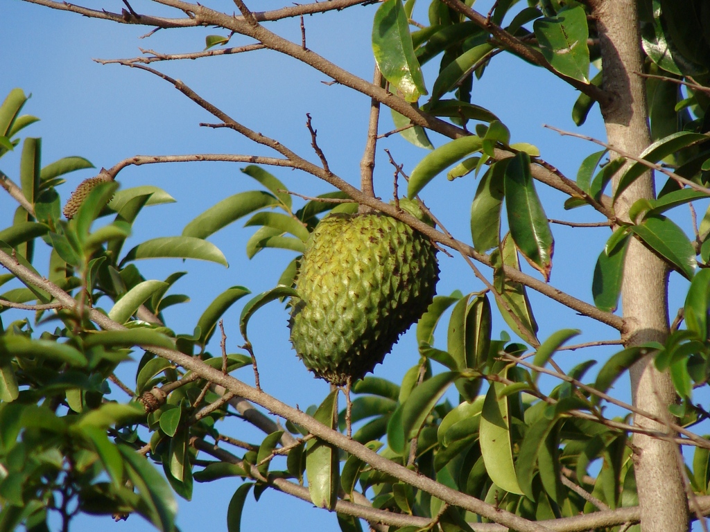
Thursday 28 March 2013

The Soursop's Anti-Cancer Properties


The Soursop Fruit on a Tree - Tatter's Photostream

Also known as the graviola, the soursop has achieved recent popularity as a cancer treatment in various alternative medicine journals.

The soursop, Annona muricata, belongs to the Annonaceae family, along with the paw paw, Asimina triloba. Initially grown for its delicious fruit, it has recently been promoted as an antic cancer treatment.
Health and alternative medicine publications continually refer to research work carried out at Purdue University under the leadership of Dr. Jerry McLaughlin, and claim that McLaughlin and his co-workers have isolated a group of chemicals from the soursop that selectively target the tumour cells of drug resistant cancers.
The Truth Behind the Anti-Cancer Claims
Mclaughlin has, in fact, carried out National Cancer Institute funded research on both the soursop and the paw paw since 1976. He has also discovered a group of chemicals known as the annonaceous acetogenins that do indeed appear to destroy tumour cells in breast, ovarian, pancreatic, liver and lung cancers. These chemicals, derivatives of long chain fatty acids, have also been shown to display pesticidal properties.
Despite these tests occurring in vitro only (that is, in the lab but not on actual cancer patients), McLaughlin’s results are encouraging. The acetogenins appear to act by inhibiting an important electron transport pathway in the production of ATP in the mitochondria of tumour cells, with the result that these cells are depleted of the energy they require to grow and multiply.
Moreover, it is the tumour cells that seem to be targeted and not normal somatic cells, as Purdue scientists discovered in tests on six human cell lines. Apparently, a special glycoprotein pump present in tumour cells that is capable of resisting the effects of anti cancer drugs is destroyed by acetogenins.
The Limited Effectiveness of the Soursop as a Cancer Treatment
Mclaughlin’s best results, however, have been with the paw paw, rather than the soursop. Two acetogenins, Bullatacin and Bullatalicin, found only in the paw paw, have proved to be much more potent than other chemicals in this family of compounds.
The Purdue team has discovered that these two molecules have double ring structures, as opposed to the less effective compounds with single or triple rings. Tests carried out by the pharmaceutical company, Pfizer, have also confirmed the effectiveness of Bullatacin in killing tumour cells.
As a consequence, while the 30 or so acetogenins in the soursop do have the capacity to destroy cancer cells, they are much less effective than those found in the paw paw. Furthermore, because extracting acetogenins from the soursop is not rigorously controlled, there is no guarantee that the concentration of this compound in the ground leaves, seeds and twigs of the plant is consistent.
Paw paw preparations, on the other hand, appear to be more stringently monitored. Paw paw twig extract, endorsed by McLaughlin, is available in capsule form from the company ‘Nature’s Sunshine’. Given the higher potency of paw paw acetogenins, coupled with its more sophisticated production techniques, it would seem that cancer patients seeking alternative treatments should choose this over any soursop products on the market.
Why Haven’t Acetogenin Treatments Been Approved by the F.D.A?
It must be noted that none of these acetogenin preparations have yet to be approved by the FDA as cancer treatments. According to the alternative medicine magazine, ‘Truth on Medicine’, large pharmaceutical companies, which could afford clinical trials, are not interested in acetogenins because they are natural compounds that cannot be synthesised.
As a result, they cannot be patented and are therefore not a profitable proposition. Mclaughlin himself adds that because the acetogenins have multiple forms (or isomers), it would be too difficult a task for these companies to process an average of around 256 possible isomers per molecule. With a range of up to 50 different acetogenins, McLaughlin believes this would be ‘simply too complex for modern studies’.
Can Acetogenins be Synthesised?
The accuracy of McLaughlin’s statement can perhaps be questioned in the light of studies by the University of Minnesota in 1993 and the Scripps Research Institute in 2000 that have in fact managed to synthesise isomers of Bullatacin, one of the most potent acetogenins. Whether this is enough for pharmaceutical companies to work with and patent is another question.
Nevertheless, the fact that independent studies continually show the tumour suppressing qualities of the acetogenins suggests a degree of truth in the claims made about the Annonaceae family. In the light of McLaughlin’s work, however, further research on the paw-paw rather than the soursop would appear to hold more promise.


References
Avedissian et.al, 2000, ‘Total Synthesis of Asimicin and Bullatacin’, The Scripps Research Institute, technion.ac.il
Cancer active, 2010, ‘Graviola anona Muricata’, canceractive.com
Fassa, P., 2010, ‘Newly Discovered Herbs Revealed for Healing Cancer and Other Diseases’, truthonmedicine.wordpress.com
McLaughlin, J., ‘Paw Paw -Alternative Cancer Treatment’ Parts 2-7, smashcancer.com
Paw Paw Research.com, 2006, ‘Graviola is Inferior to Paw-Paw as a Cancer Treatment: Here’s why’, pawpawresearch.com


Vitamins and the Evolution of Skin Colour


Figure 1:Darwin Meets NASA
Nina Jablonski - NASA
Current research suggests the global distribution of skin colour has arisen through an evolutionary process involving UV radiation and vitamins.

According to studies made by the State University of New York College in 1997, the global distribution of skin colouration can be correlated with UV radiation levels. To be more precise, amounts of melanin in the skin were found to be highest at the equator. They then decreased by about 8% for each 10 degrees of latitude in the Northern Hemisphere and by about 4% for each 10 degrees of latitude in the Southern Hemisphere.
Darwin and Natural Selection
Many scientists believe that this range of skin colouring has arisen as a result of evolution. The fundamental idea behind the theory of evolution is that all the species alive on Earth today have descended from a simple common ancestor. In his Origin of Species, published in 1859, Charles Darwin proposed a mechanism for this, called ‘Natural Selection’.
This theory effectively states that in the struggle for existence, only those individuals that are adapted to their surroundings will survive to reproduce. The favourable genetic characteristics that allowed them to survive will then be passed on to their offspring.
How Does Natural Selection Explain Skin Colour?
When contemplating the evolution of skin colour, an initial hypothesis might be that dark skin predominates in equatorial areas because people with light skin were more susceptible to skin cancer. The flaw in this idea, of course, is that most people develop skin cancers later in life, well after they have reproduced and passed their genes for fair skin to their offspring.
Moreover, it doesn’t explain why fair skinned people predominate in northern latitudes. The amount of UV radiation is markedly reduced here, so why should humans with any skin colour, let alone dark skinned people, have died out or been prevented from reproducing in these areas? The answer, according to anthropologist Nina Jablonski , is that deficiencies in Vitamin B folate (folic acid) and vitamin D, rather than skin cancer, have been the evolutionary drivers of skin pigmentation.
Fair Skin and Folate Deficiency
In studies made in 1978 Jablonski found that excessive exposure to UV rays destroys blood folic acid. In equatorial regions, fair skinned people lacking sufficient melanin to block UV radiation may consequently have been at risk of having reduced levels of this vitamin.
Because folic acid is important in embryonic neural tube formation, Jablonski concludes that natural selection favoured people with dark pigmentation in these areas because they could produce healthy offspring. Moreover, a lack of folate reduces sperm production and DNA synthesis, which may have further contributed to the demise of fair skinned people in warm climates.
Dark Skin and Vitamin D Deficiency
Conversely, people with fair skin may be at an advantage in more extreme latitudes where UV radiation is weaker. This is because UV B light is needed to manufacture vitamin D3, important in calcium absorption. Vitamin D is particularly vital during pregnancy for depositing calcium in the fast growing bones of the foetus, and also for foetal growth and development of the nervous system, lungs and immune system.
Jablonski hypothesises that this need for vitamin D during pregnancy may be why women in fact tend to have lighter skin than men, and why reproduction may not have been as successful in darker skinned women in northern latitudes.
Diet and Vitamin D Deficiency
According to some researchers, UV radiation as a source of vitamin D may only have increased in importance over the last 10,000 years. Prior to this, when humans were predominantly hunter-gatherers, vitamin D was also readily available in the meat and fish prevalent in their diet. Indeed, the Inuit Indians, whose diet includes ocean fish, seem to have evolved successfully in Arctic Regions despite having dark skins.
Heather Norton, a molecular anthropologist at the University of Arizona, has made studies of the mutant SLC24A5 gene, responsible for fair skin pigmentation, and agrees with the proposition that it only appeared around 6000 years ago. Careful analysis revealed little to no variation in the DNA surrounding this gene, suggesting that less time had passed since it appeared than previously believed.
Is Vitamin D a Genuine Selecting Agent?
With the introduction of agriculture and sedentary lifestyles, then, dark skinned people must not have received sufficient Vitamin D to successfully reproduce, leading to their ultimate decline in northern latitudes. Interestingly, however, researchers at the University of Wisconsin have found that rats deprived of Vitamin D are still capable of reproducing, albeit with the production of smaller litters and decreased neonatal growth rates.
Is this enough, then, to result in the disappearance of all dark skinned people in extreme latitudes? Perhaps not, but according to researcher Reinhold Vieth, pelvic deformity in women with rickets (a disease caused by Vitamin D deficiency) also makes a vaginal delivery virtually impossible. This would have further decreased the numbers of offspring produced by dark skinned women.
Nina Jablonski’s theory certainly holds some merit, and remains the most logical explanation for the evolution of skin colour throughout the globe. As Jablonski herself states, perhaps Darwin himself would have agreed with her if he’d had access to such modern technology as NASA’s TOM’s satellite (see figure 1), which has recorded the global distribution of UV radiation. At the very least, it may have inspired him to discuss human evolution, a subject that he only briefly touched on in his publications.
References
Halloran, B. and De Luca, H.,1980, 'Effect of Vitamin D Deficiency on Fertility and Reproductive Capacity in the Female Rat', nutrition.org
Jablonski, N. and Chaplin, G., 2000, 'The Evolution of Human Skin Coloration', California Academy of Sciences, yanaiweb.com
Jablonski, N., 2009, 'Jablonski Breaks the Illusion of Skin Colour', ted.org
Norton, H., Hammer, M., 'Sequence Variation in the Pigmentation Gene SLC24A5... and the evolution of Light Skin', physanth.org
Relethford, j.H., 1998, 'Hemispheric Difference in Human Skin Colour', Am J Phys Anthropol, nih.gov
Vieth, R., 'Effects of Vitamin D on Bone and Natural Selection of Skin Colour; How much Vitamin D Nutrition Are We Talking About?' ms.org


Evolution of the Hippopotamus


The Pygmy Hippo
Pygmy Hippo - Zoe Bianchi
The Hippopotamus, an even-toed ungulate, is now believed to be more closely related to whales than to pigs and peccaries.
The pygmy hippopotamus and the common or Nile hippopotamus are thought to have evolved from a common ancestor that lived some 10 million years ago. This common ancestor belonged to a group of creatures known as anthracotheres – semi-aquatic even-toed ungulates belonging to the order Artiodactyla. The hippo is placed the same order as giraffes, cattle, goats, pigs, camels and peccaries.

The Relationship Between Hippos, Anthracotheres and Whales
This proposition has been supported by fossil comparisons, which show similarities between the teeth and skulls of anthracotheres and the hippopotamus. For the past 20 or more years, however, molecular studies have also suggested a relationship between the hippo and whales. This has been something more difficult to determine using fossil evidence, as the two groups superficially bear no physical resemblance.
Recent discoveries of ancient whale remains in Pakistan, however, have finally made a physical connection between cetaceans (whales and dolphins) and artiodactyls. In 2000, Professor Philip Gingerich and his co-workers uncovered the 47 million year old remains of a whale ankle bone that clearly placed it in the Artiodactyl order. The overall structure of the ‘leg’ bones of the whale also suggested that it could use them to prop itself up on land for brief periods.

Indohyus and Raollids
This finding, combined with immunological, blood protein and DNA evidence (due to the work in 1985 of Vincent Sarich, of Berkely University), appears to place the hippopotamus as the closest living relative to the whale. The fossil species Indohyus, an even–toed ungulate resembling a small deer, has even more in common with whales, possessing structural similarities in the skull and ear regions.
A research team led by Dr. Hans Thewissen has also concluded from bone density and dental studies that Indohyus was aquatic. The fact that Indohyus (a member of the extinct order Raoellidae) was a vegetarian has also led them to surmise that the carnivorous nature of whales evolved after they returned to the water.
Jean-Renaud Boisserie and colleagues at the University of California believe the anthracotheres to be a common ancestor of both the hippos and whales. Thewissen and co workers , however, prefer the idea that the ancestors of whales (which include Indohyus and other Raollids) belong to a side group that branched away from other artiodactyl groups before the anthracotheres appeared.
How Closely Related Are Hippos, Horses and Pigs?
At any rate, the ancestry of the hippopotamus seems clearer. Although the word ‘hippopotamus’ is derived from the Greek word meaning ‘river horse’, there is no direct relationship between horses and the hippopotamus, as the ancient Greeks believed. Horses in fact belong to the Perissodactyla order, consisting of odd-toed ungulates with more simplistic digestive systems than the artiodactyls. Other perissodactyls include the rhinoceros and the tapir.
While pigs and peccaries are indeed artiodactyls, they are not as closely related to the hippopotamus as scientists have previously thought. Most current researchers now combine DNA and fossil evidence to derive evolutionary relationships between different species. As a result, most, including Jessica Theodor and Jonathan Geisler of the University of Calgary, have concluded that hippos are in fact more closely related to whales than to pigs.
The Relationship Between the Pygmy Hippo and the Common Hippo
Pygmy Hippo
Pygmy Hippo
As members of the Artiodactyl order, the common Hippopotamus, Hippopotamus amphibious and the Pygmy Hippopotamus, Hexaprotodon liberiensis, bear strong anatomical similarities to each other, despite the fact that the pygmy hippo is believed to have branched off from the common hippo over 10 million years ago.
Although both are aquatic, the pygmy hippo is nocturnal and less gregarious than the Nile Hippo, and possesses less webbing between the toes. It is also much smaller in size (having a mass of between 180-720 kg as opposed to up to 2000 kg in the case of the common hippo), with a different diet.
Pygmy hippos are in fact threatened with immediate risk of extinction, due to a combination of hunting and habitat destruction in Sierra Leone and other areas of Western Africa. Thankfully, they are breeding quite successfully in captivity, which may help to ensure these unusual artiodactyls, along with the common hippos, remain with us for a little longer.
Sources
                Arbor, A., 2001, ‘New Fossils Suggest Whales and Hippos are Close Kin’, ScienceDaily.com. Accessed May 15, 2011.
                Celeskey, M., 2010, ‘Indohyus and Cetacean Relations’, VladimirKorsakov.Blogspot.com. Accessed May 15, 2011.
                Robinson, P.T., ‘River Horses and Water Cows’, University of California, San Diego, ML.Duke.edu. Accessed May 15, 2011.
                Sanders, R., 2005, UC Berkeley, French Scientists Find Missing Link Between the Whale and its Closest Relative, the Hippo’, ScienceDaily.com. Accessed May 15, 2011.


Sunday 24 March 2013

Parkinson's Disease and Stem Cells


The Brain, American Museum of Natural History
Human Brain - Gasz's Photostream

Parkinson's disease, caused by the degeneration of dopamine producing cells, may be among the first conditions to be successfully treated using stem cells.

Parkinson’s disease is a condition in which the neuronal message to the muscles is disrupted, resulting in symptoms such as tremors and the impairment of movement and speech. It is caused by the degeneration of dopamine producing cells in an area of the mid region of the brain known as the substantia nigra.
Dopamine is a neurotransmitter – a molecule capable of converting nervous impulses to chemical messages across the synapses, or gaps, between one neurone and the next. It is associated with the control of movement, emotional responses and our ability to experience pleasure and pain.
The Benefits of Stem Cells Compared to Drugs
Various drugs such as Levodopa, Sinemet and the dopamine agonists Requip and Mirapex can either act to initiate more dopamine production or mimic the action of dopamine. However, they ultimately have only limited effectiveness, can cause side effects such as dyskinesias (uncontrolled movements), nausea and confusion and do not reduce the degeneration of the dopamine producing (dopaminergic) nerve cells themselves.
A better and longer lasting treatment would ideally be one where the dopaminergic cells are replaced with healthy ones that can regenerate themselves over the course of the patient’s life. Stem cell therapy, in which differentiated stem cells of embryos or modified adult stem cells, are implanted into the brain, may offer such a solution.
The Current State of Stem Cell Trials
To date, approved scientific trials have only involved the introduction of stem cells to ‘models’ of Parkinson’s disease rather than to human patients themselves. This has usually involved modifying the metabolism of laboratory rats to mimic the symptoms of the disease and then injecting stem cells that have been manipulated into forming dopaminergic cells into the rats.
Researchers at the Michael J. Fox Foundation have found that such trials have been the most successful when embryonic stem cells have been used. It is for this reason that this organization, among others, has campaigned for federal funding for stem cell research since 2001. Although limited funding has been approved over the years, there are still restrictions on the stem cell lines that are available for research purposes.
Induced Pluripotent Stem Cells (iPSCs) and the Future
Fortunately, an alternative to the use of embryonic stem cells was discovered in 2007. Researchers at the University of Wisconsin and the University of Kyoto concurrently found that adult human fibroblast cells, commonly found in connective tissue, could be genetically altered to produce the same proteins as embryonic stem cells. This allowed them to become ‘pluripotent’: in other words, capable of differentiating into a variety of specialized cells, including dopaminergic ones. Other research, involving the reprogramming of neural and testicular cells into pluripotent cells, is also currently underway.
Initially, this modification process involved the introduction of genes to the fibroblast cells via a retrovirus. However, the safety and efficacy of this method have been questioned by scientists at the Whitehead Institute and the Harvard Medical School, who have noted that viral vectors can sometimes affect the differentiation potential of fibroblasts or even cause malignant cell growth.
One solution, they claim, is the use of ‘factor free’ iPSCs – induced fibroblast cells in which the viral reprogramming factors have been removed. This involves the use of an enzyme called Cre-recombinase which helps to rid the iPSCs of potentially harmful transgenes.
Protein Based iPSCs
Alternatively, Sang-Hun Lee, at Hanyang University, Korea, and Kwang-Soo Kim, at Harvard Medical School have developed iPSCs in which the required reprogramming proteins are themselves introduced into fibroblast cells, thus obviating the need for virus delivered transgenes. When these ‘protein based’ iPSCs were implanted into rats modeling Parkinson’s disease the symptoms appeared to be alleviated.
Whether or not any one particular method is more successful than the other, it appears clear that iPSCs are offering new hope and a possible alternative to the ethical issues surrounding the use of embryonic stem cells. Recent tests at the Universities of Göttingen and Tübingen have, moreover, shown that iPSC derived dopaminergic cells can be delivered to rats via nasal sprays rather than by injections. Scientists involved with this research found that the cells became successfully incorporated into the rat brain tissue, surviving and producing dopamine for up to six months.
The Continuing Need for Both Types of Stem Cells
Despite the moderate success of iPSCs, researchers are in agreement that funding for embryonic stem (ES) cell research needs to continue, if only so that the effects of iPSCs can be regularly compared to ES cells in the lab. According to University of Michigan sociologist Owen Smith, 62.1% of current scientific papers on stem cell research involved using both iPS and ES cells together.
At any rate, apart from the complexities of manipulating stem cells to form dopaminergic cells, there are still questions regarding the quality and quantity of dopamine production by these cells if, and when, they are incorporated into Parkinson’s patients. Immune rejection, even of dopaminergic cells derived from the patient’s own body, remains a distinct possibility. In the words of famous Parkinson's sufferer, Michael J. Fox, the struggle for a cure for this disease constantly involves ‘one step forward and two steps back.’
References
                Euro Stem Cell, 2010, ‘What is Parkinson’s Disease and Can Stem Cells Help?’ eurostemcell.org
                Journal of Clinical investigation, May 2011, 'Stem Cells May Reverse Parkinson's Disease', Ivanhoe.com
                Kiessling, A., 2010, ‘The State of the Stem Cell’, bedfordresearch.org
                Michael J. Fox Foundation for Parkinson's Research, Research Area Position Papers, 2009, ‘Stem Cells 101’, michaeljfox.org
                Science Daily, 2011, ‘Dramatic Improvement in Parkinson Disease Symptoms Following Intranasal Delivery of Stem Cells to Rat Brains’, sciencedaily.com
                Soldner et. al, 2009, Whitehead Institute, Harvard Medical School, ‘Parkinson's Disease Patient-Derived Induced Pluripotent Stem Cells Free of Viral Reprogramming Factors’, cell.com


Friday 22 March 2013

Classifying Primates


Philippine Tarsier - Roberto Verzo's Photostream
The latest trend in primate classification has been to classify in a manner that best reflects evolution, geography and physical and biochemical factors.
Primates are mammals possessing a generally accepted group of physical characteristics. These include opposable thumbs, a relatively large brain, eyes that are oriented towards the front of the head, two nipples and stereoscopic (binocular) vision.
Until recently, taxonomists have generally adopted the similar classification systems proposed by Simons in 1972 and Fleagle in 1998. According to these systems, the primate order was divided into two sub-orders: the prosimians and the anthropoids.
Prosimians included lemurs, lorises and tarsiers, grouped together because they were considered morphologically more primitive than other primates. The anthropoids comprised all other primates – in other words, monkeys, apes and man.
Fleagle’s system also placed humans in the superfamily Hominoidea, along with apes. The apes, which included orangutans, gibbons, gorillas, chimpanzees and bonobos, were then separated into the family Pongidae, while humans were grouped into the Hominidae.
The Re-Classification of Tarsiers
Recent anatomical and biochemical studies, however, have thrown some doubt on this system. According to Ben F. Koop and fellow researchers at the Wayne State University, for instance, the alpha and beta globin genes in tarsiers (see fig.1) bear more resemblance to those of the anthropoids than those of the prosimians. DNA hybridization comparisons and RNA sequencing have also suggested a similar link.
In addition, the nostrils of tarsiers and anthropoids are surrounded by dry skin whereas lemurs and lorises have moist nostrils. Other physical differences, such as the absence of a tapetum (a layer in the retina that helps in night vision) in tarsiers and anthropoids, have led many taxonomists to devise two new primate sub-orders. These are the Strepsirhines (lemurs and lorises) and the Haplorhines (tarsiers, monkeys, apes and humans).
In evolutionary terms, this new grouping coincides with the view that tarsiers represent an evolutionary bridge between the two groups (see fig.2) and are in fact quite unique in many aspects of their morphology and behaviour. Jason Cowan, of Washington State University goes so far as to suggest they should be placed in a sub-order of their own.
New Groupings for Apes and Humans
Moreover, molecular comparisons of members of the super family Hominoidea have revealed that gorillas and chimpanzees are more closely related to humans than other apes. This similarity is so pronounced that they have now been placed together with humans in the family Hominidae. In fact, 98.77% of the DNA in humans and chimpanzees is identical, representing an evolutionary divergence that occurred as recently as 6 million years ago.
Gibbons, on the other hand, have been grouped with siamangs into the family Hylobatidae. According to Evans, 2009, this family represents a closer evolutionary link to Old World Monkeys (a 27 gene difference) than the Hominidae (a 31- 36 gene difference) and the Pongidae (a 33 gene difference). The Pongidae family now consists only of the orangutans, a group of South East Asian apes that diverged from other hominoids around 11 million years ago.
Note that another system places the orangutans in the Ponginae, a sub-family of the Hominidae, as a result of their close genetic relationship to this family. The African apes (gorillas, chimps and bonobos), however, remain the nearest evolutionary link to humans.
Geographical Distribution and Evolution
Considering humans are believed to have arisen in Africa, our close relationship to the African apes appears to make sense. Humans and apes in fact belong to the infra-order catarrhines because, along with biochemical similarities, they have downward pointing noses and lack prehensile (branch-gripping) tails. Old World Monkeys such as baboons and macaques are also included in this group, and can be found in Africa or Asia.
Similarly, New World monkeys (infra-order platyrrhines), all found in South America, Central America and the Caribbean islands, are grouped together because of physical similarities such as broad noses and prehensile tails. Lemurs and lorises are found only in Madagascar, while tarsiers are limited to a narrow geographical range within South East Asia.
These findings are consistent with the concept that evolution occurs in populations that are geographically isolated. The Strepshirines (lemurs and lorises) for instance, are a unique group of primates largely because Madagascar, isolated from India for over 80 million years and separate from the coast of Africa, has provided ideal conditions for natural selection to have occurred.
Similarly, the New World Monkeys may have evolved to develop their particular features after South America separated from North America. Alternatively, according to McKenna (1980), they possibly arrived some time later by ‘island’ hopping from North America via the Caribbean islands. In any case, the original primates to arrive in this continent had ample time to evolve in relative isolation.
It would appear, then, that geographical distribution and evolutionary trends, along with physical and biochemical factors, all tend to support current primate classification systems. The creation of the new sub-orders, Strepshirines and Haplorhines, to reflect the unique nature of the Tarsier, is perhaps the best example of this.
References
                Cowan, J., Washington State University, 2006, 'Are Tarsiers Really a Taxonomic Enigma?', soar.wichita.edu
                Fleagle, 1998, 'Taxonomy of the Primates', theprimata.com
                McKenna,M (1980), 'Early History and Biogeography of South America's Extinct Land Mammals', Plenum, New York
                Simons, E. 1972, 'Primate Evolution', Macmillan, New York